Commonly referred to as B (brightness) mode, the use of grey scale imaging in ultrasound renders a two-dimensional image in which the organs and tissues of interest are depicted as points of v
Grey-scale and Doppler ultrasound. Transverse (a) and sagittal (b) grey
Diaphragmatic hernia - Radiology at St. Vincent's University Hospital
Grey scale imaging (ultrasound), Radiology Reference Article

Utility of Ultrasound in the Diagnosis, Treatment, and Follow-up of Prostate Cancer: State of the Art
On the superior panel, 2D gray scale sonography demonstrates a

Role of gray-scale and color Doppler ultrasound in women with chronic pelvic pain Abdullah MS, Mousa WA, Ghobashy MA - Menoufia Med J
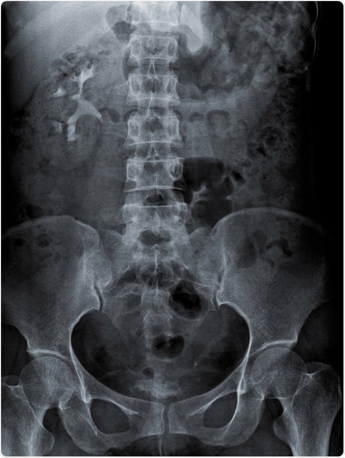
KUB Radiography

PDF] From Grey Scale B-Mode to Elastosonography: Multimodal Ultrasound Imaging in Meningioma Surgery—Pictorial Essay and Literature Review
CT of Calcifying Jaw Bone Diseases
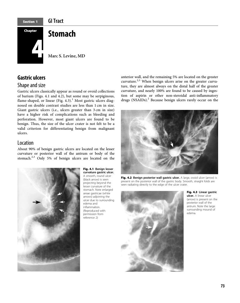
Stomach (Chapter 4) - Practical Fluoroscopy of the GI and GU Tracts

PDF] From Grey Scale B-Mode to Elastosonography: Multimodal Ultrasound Imaging in Meningioma Surgery—Pictorial Essay and Literature Review
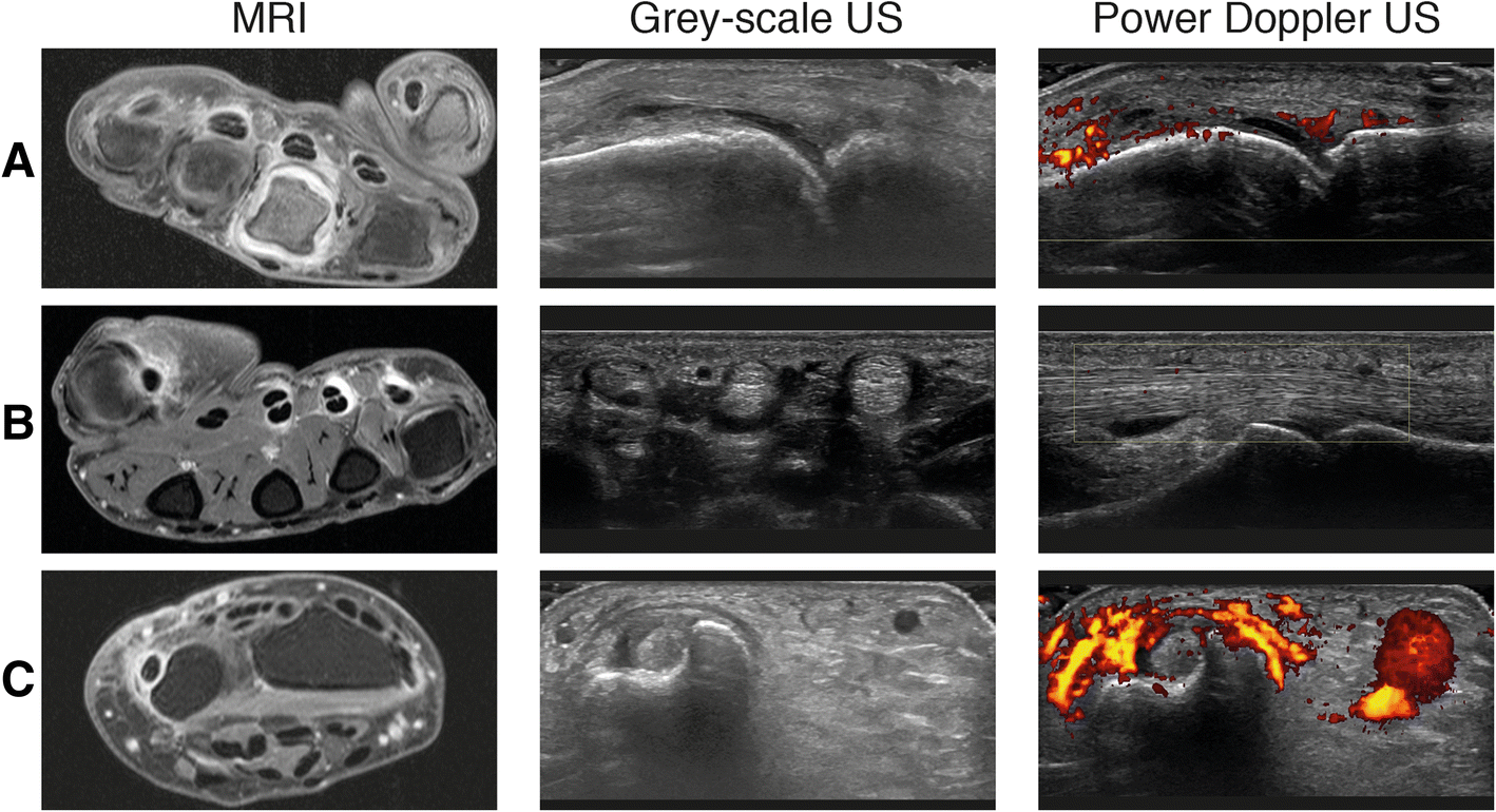
Do musculoskeletal ultrasound and magnetic resonance imaging identify synovitis and tenosynovitis at the same joints and tendons? A comparative study in early inflammatory arthritis and clinically suspect arthralgia

What do we know about volumetric medical image interpretation?: a review of the basic science and medical image perception literatures, Cognitive Research: Principles and Implications
XDR Radiology Releases XDR v3.2.11 - XDR Radiology