Schematic depiction of the distribution of the PV autoantigens Dsg1

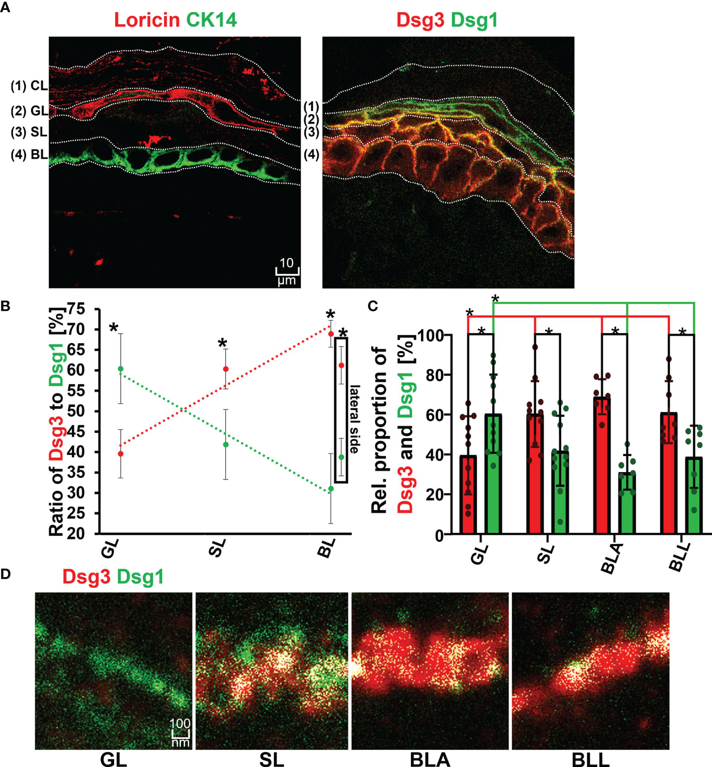
Download scientific diagram | | Schematic depiction of the distribution of the PV autoantigens Dsg1 (green) and Dsg3 (red) and the composition of desmosome along different epidermal layers in normal epidermis (left) and PV-affected epidermis (right). *Significant difference to the value which is indicated that it is compared to. from publication: Dsg1 and Dsg3 Composition of Desmosomes Across Human Epidermis and Alterations in Pemphigus Vulgaris Patient Skin | Desmosomes are important epidermal adhesion units and signalling hubs, which play an important role in pemphigus pathogenesis. Different expression patterns of the pemphigus autoantigens desmoglein (Dsg)1 and Dsg3 across different epidermal layers have been demonstrated. | Desmosomes, Pemphigus and Epidermis | ResearchGate, the professional network for scientists.

Dsg1 and Dsg3 Composition of Desmosomes Across Human Epidermis and Alterations in Pemphigus Vulgaris Patient Skin. - Abstract - Europe PMC

Jens WASCHKE, Ludwig-Maximilians-University of Munich, München, LMU, Institute for Anatomy and Cell Biology

PDF) Dsg1 and Dsg3 Composition of Desmosomes Across Human Epidermis and Alterations in Pemphigus Vulgaris Patient Skin

Structural changes of human keratinocytes in response to pemphigus

Daniela KUGELMANN, Ludwig-Maximilians-University of Munich, München, LMU, Faculty of Medicine

Single-Cell Transcriptomes and Immune Repertoires Reveal the Cell State and Molecular Changes in Pemphigus Vulgaris

Schematic depiction of the distribution of the PV autoantigens Dsg1
Frontiers Dsg1 and Dsg3 Composition of Desmosomes Across Human Epidermis and Alterations in Pemphigus Vulgaris Patient Skin

Immunogenetic mechanisms for the coexistence of organ-specific and systemic autoimmune diseases, Journal of Autoimmune Diseases

Daniela KUGELMANN, Ludwig-Maximilians-University of Munich, München, LMU, Faculty of Medicine