Medial view of left knee region highlighting various fascial

Download scientific diagram | Medial view of left knee region highlighting various fascial components surrounding the semitendinosus muscle. From the superficial to the deep aspect: the fascia lata, the paratenon and the epimysium from publication: Anatomical study of paratenons and fascia lata connections in the posteromedial knee region | Introduction In the last decade, fascia research increased significantly in various aspects such as anatomical and biomechanical features related to epimuscular force transmission. Methods The present anatomic study focuses on macroscopic observations of the potential | Fascia Lata, Hamstring muscles and Fascia | ResearchGate, the professional network for scientists.
Scrotum: Anatomy, blood supply, innervation and function

Sharing of proximal fibers by the anterolateral and lateral
Superior tibial plateau view of the Medial and Lateral Meniscus as well as the anterior and posterior cruciate ligament. Bent knee view of the right

Normal Left Knee Anatomy - Superior tibial plateau, sagittal & anterior view

Influence of different knee and ankle ranges of motion on the
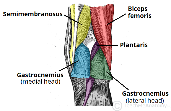
The Popliteal Fossa - Borders - Contents - TeachMeAnatomy
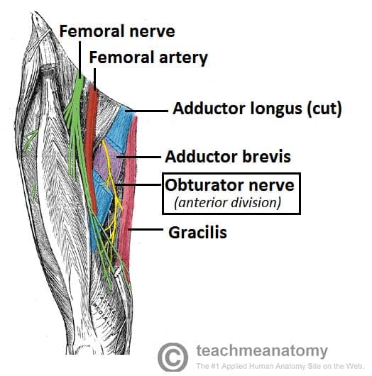
The Obturator Nerve - Course - Motor - Sensory - TeachMeAnatomy

Knees: How Scott free Somatics, a gentle method of working with

Knee Self Treatment Taylor Made Integrative Therapy

Medial and Anterior Knee Anatomy

Medial view of right knee region highlighting gracilis (G) and