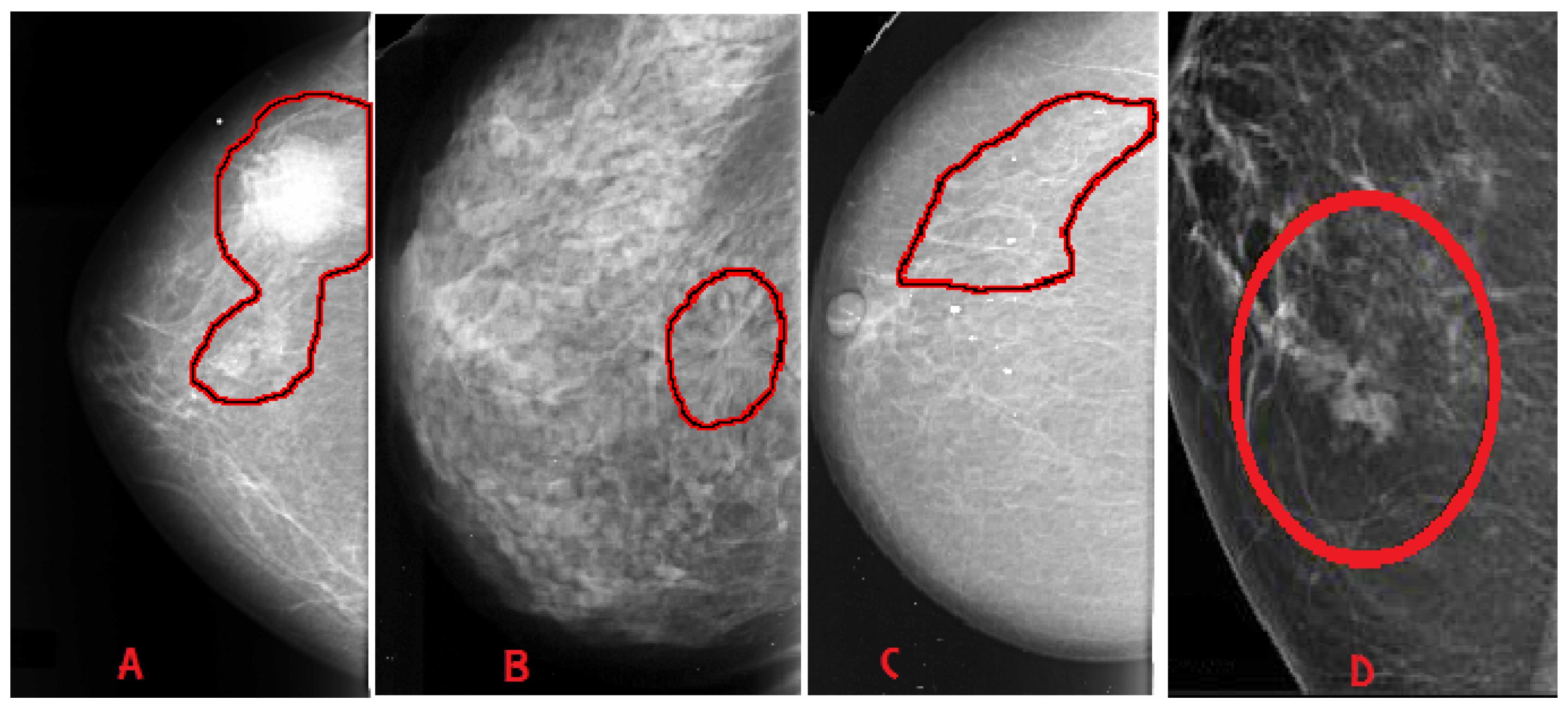
Calcification and mass abnormalities in breast mammogram scans
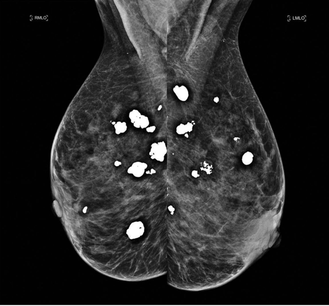
Download scientific diagram | Calcification and mass abnormalities in breast mammogram scans. The calcification distribution depicts tiny flecks of calcium as small white regions on the left side, while the mass is shown as a smooth, well-defined border on the right side. from publication: Multi-Graph Convolutional Neural Network for Breast Cancer Multi-Task Classification | Mammography is a popular diagnostic imaging procedure for detecting breast cancer at an early stage. Various deep learning (DL) approaches to breast cancer detection incur high costs and are prone to classify incorrectly. Therefore, they are not sufficiently reliable to | Breast Cancer, Convolution and Classification | ResearchGate, the professional network for scientists.
:max_bytes(150000):strip_icc()/lateral-mammogram-of-female-breast-with-tumor-92263689-813095ee469b45eabfc9f5f4747758ed.jpg)
Mammogram Images: Normal and Abnormal
J. Imaging, Free Full-Text
What does pleomorphic calcifications mean on a mammogram? What about focal asymmetry with architecture distortion? I had a screening mammogram and am being called back for additional imaging. Is this something I
Contrast enhanced mammography: focus on frequently encountered benign and malignant diagnoses, Cancer Imaging

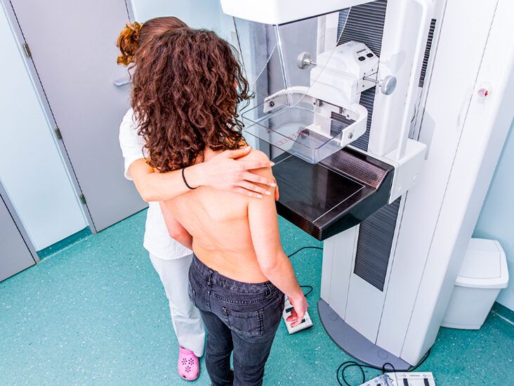
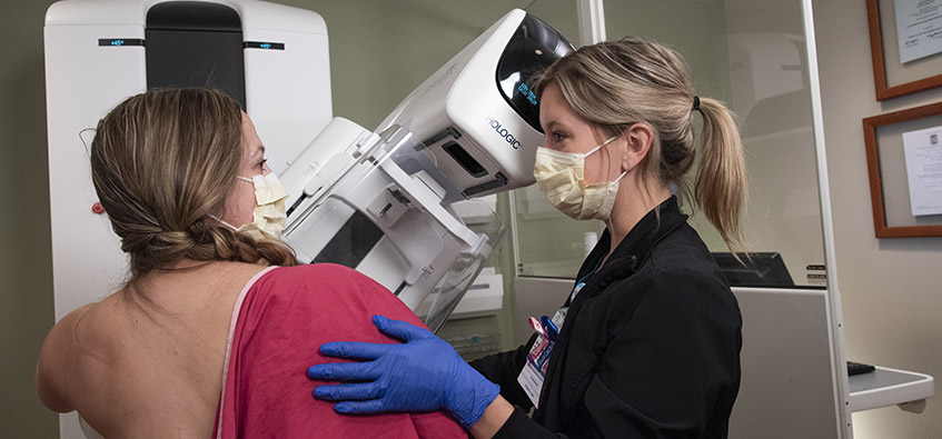
What Do Spots on a Mammogram Mean? - Health Images

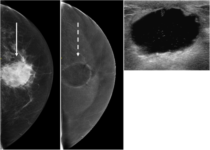
Contrast enhanced mammography: focus on frequently encountered benign and malignant diagnoses, Cancer Imaging
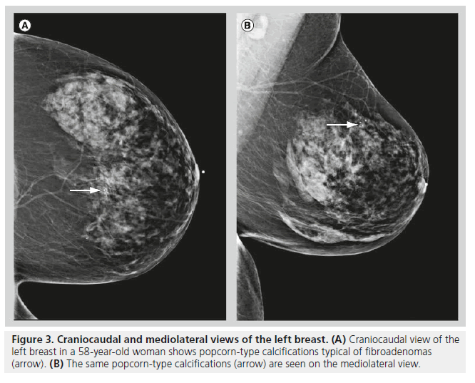
Mammography of breast calcifications
Mammography: Masses - Radiology

Breast Lesions Detection and Classification via YOLO-Based Fusion Models
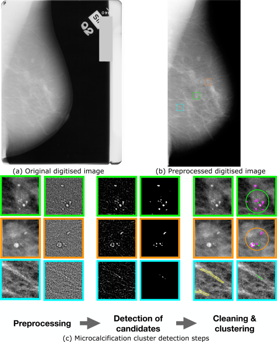
Association of Microcalcification Clusters with Short-term Invasive Breast Cancer Risk and Breast Cancer Risk Factors

A 58-year-old female with complaints of skin discoloration and vague
Atlas of breast cancer early detection

PDF) Multi-Graph Convolutional Neural Network for Breast Cancer
Breast calcifications mimicking pulmonary nodules