Cellular fluorescence intensity and cell size as a function of

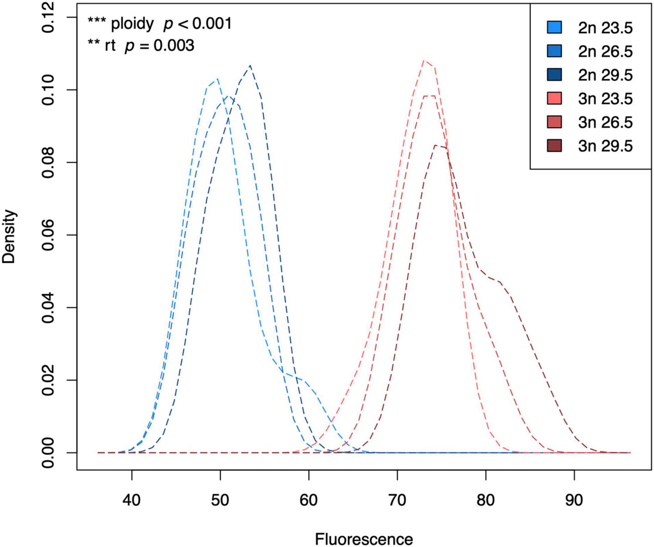
Download scientific diagram | | Cellular fluorescence intensity and cell size as a function of enrichment substrate on a subset of basalt enrichments (sample 30R-1A). The circle size indicates the average cellular area (mm 2 ) as measured by epi-fluorescence microscopy. The standard deviation for the cellular area of each sample is roughly 46% based on the following number of cells counts for each treatment: Host Rock (n = 28); No addition (n = 41); CH 3 COONa (n = 38); CH 4 (n = 62); NaHCO 3 (n = 43); NaHCO 3 + NH 4 Cl (n = 39); NaHCO 3 + NaNO 3 (n = 36); NH 4 Cl (n = 35); NaNO 3 (n = 38). Symbols ++ and + indicate analysis of variance P-values of <0.01 and <0.05, respectively, versus Host rock or No addition; and * * indicate P < 0.01 versus CH 3 COONa, CH 4 or NaHCO 3 . from publication: Nitrogen Stimulates the Growth of Subsurface Basalt-associated Microorganisms at the Western Flank of the Mid-Atlantic Ridge | Oceanic crust constitutes the largest aquifer system on Earth, and microbial activity in this environment has been inferred from various geochemical analyses. However, empirical documentation of microbial activity from subsurface basalts is still lacking, particularly in the | Nitrogen, Stimulation and Geomicrobiology | ResearchGate, the professional network for scientists.

Cell size homeostasis is maintained by CDK4-dependent activation of p38 MAPK - ScienceDirect

Variability of fluorescence intensity distribution measured by flow cytometry is influenced by cell size and cell cycle progression

PDF) Nitrogen Stimulates the Growth of Subsurface Basalt

PDF) Nitrogen Stimulates the Growth of Subsurface Basalt

A) The percentage of positive cells, (B) mean fluorescence intensity
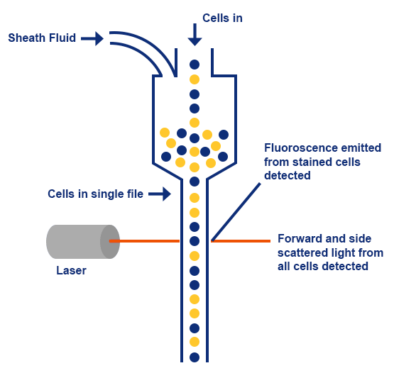
Guide to Fluorescence Activated Cell Sorting (FACs): The Latest Technology in Cell Separation - Assay Genie

PDF) Nitrogen Stimulates the Growth of Subsurface Basalt
Frontiers Interacting Effects of Cell Size and Temperature on Gene Expression, Growth, Development and Swimming Performance in Larval Zebrafish
Frontiers The Application of Imaging Flow Cytometry for Characterisation and Quantification of Bacterial Phenotypes

Characteristics of IODP Expedition 336 samples used in this study

Characteristics of IODP Expedition 336 samples used in this study

Number of cells versus the fluorescence intensity histogram. The first

Fast and accurate automated cell boundary determination for fluorescence microscopy

Schematics of SAPS (successive alkalinity producing system
Abbexa Ltd - Flow Cytometry