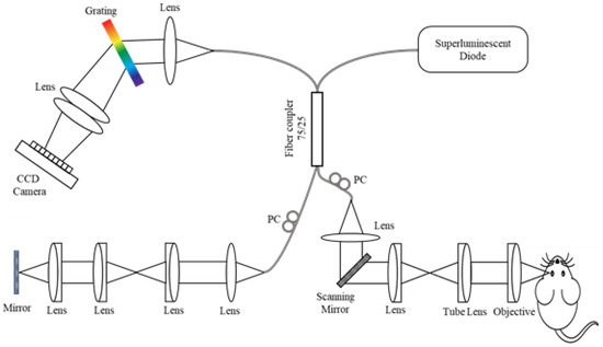
Optical Coherence Tomography: Imaging Mouse Retinal Ganglion Cells In Vivo

Scientific Article | Structural changes in the retina are common manifestations of ophthalmic diseases.
Durable 3D murine ex vivo retina glaucoma models for optical coherence tomography
Applied Sciences, Free Full-Text

Jolanta JAGODZINSKA, PhD Student, Master of Science, Institut des Neurosciences de Montpellier, Montpellier, INM, Vision

Optical coherence tomography angiography (OCT-A) in an animal model of laser-induced choroidal neovascularization - ScienceDirect

Fig. 9.11, [In vivo confocal reflectance and]. - High Resolution Imaging in Microscopy and Ophthalmology - NCBI Bookshelf
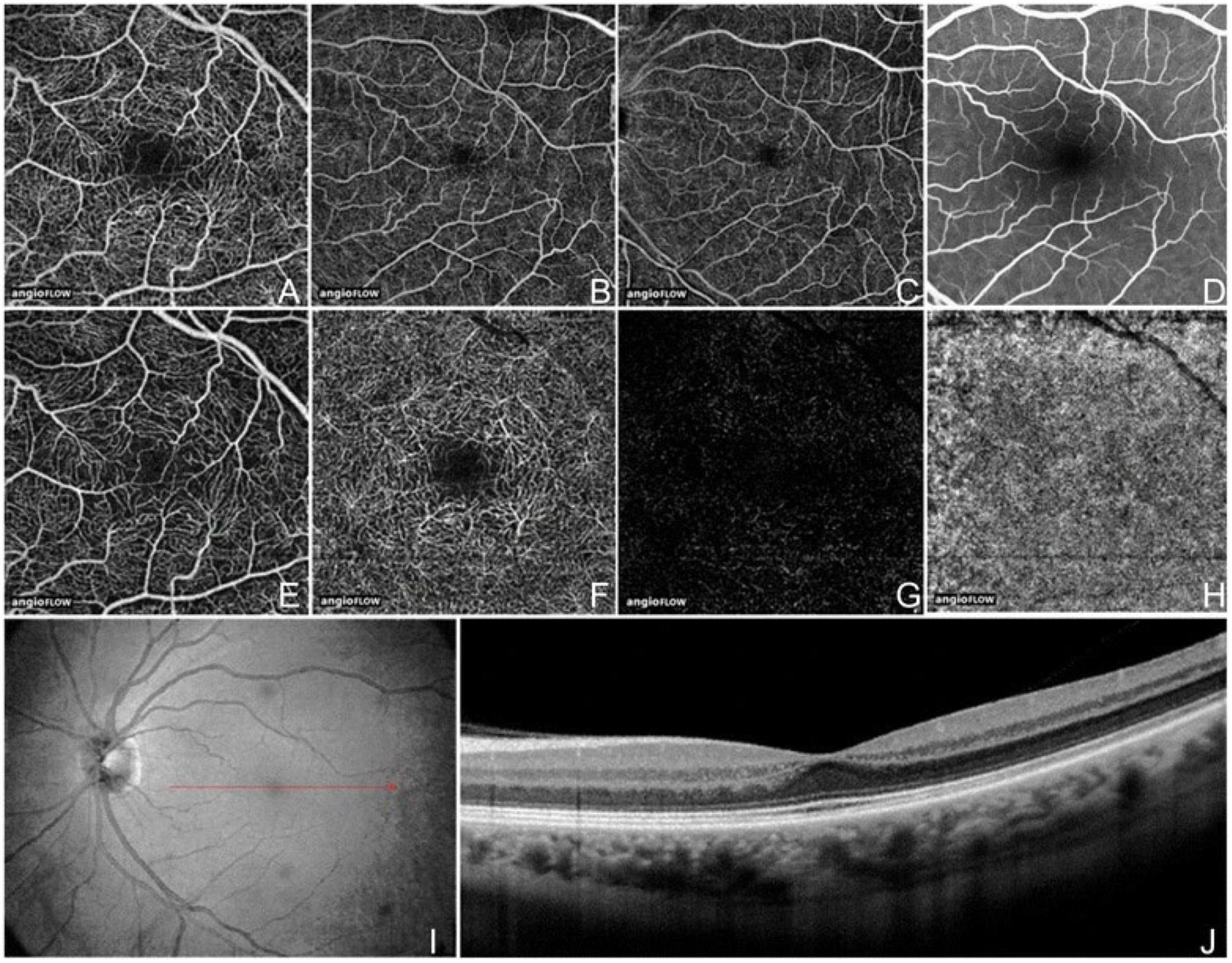
Frontiers The Development and Clinical Application of Innovative Optical Ophthalmic Imaging Techniques

Image-Guided Optical Coherence Tomography to Assess Structural Changes in Rodent Retinas

Genes, Free Full-Text

In vivo imaging of mouse retina. The spectral domain-optical

Fig. 9.4, [In vivo CSLO images of]. - High Resolution Imaging in Microscopy and Ophthalmology - NCBI Bookshelf

Topical Nerve Growth Factor (NGF) restores electrophysiological alterations in the Ins2Akita mouse model of diabetic retinopathy - ScienceDirect

Correction-free remotely scanned two-photon in vivo mouse retinal

PDF] Quantitative Analysis of Mouse Retinal Layers Using Automated Segmentation of Spectral Domain Optical Coherence Tomography Images.

Human adipose tissue-derived stem cell extracellular vesicles attenuate ocular hypertension-induced retinal ganglion cell damage by inhibiting microglia- TLR4/MAPK/NF-κB proinflammatory cascade signaling, Acta Neuropathologica Communications

Christian P Hamel's research works Université de Montpellier, Montpellier (UM1) and other places